Research
Cellular and molecular biology of the implanted human cochlea
We are investigating the effects of the cochlear implant (CI) in the different cell types of the human inner ear using immunohistochemistry.
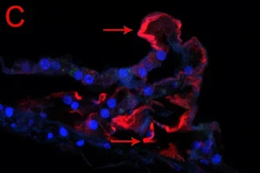
- We are investigating the fate of the neurons and the remaining cells of the organ of Corti using specific cellular markers. We are using antibodies against neurofilaments and acetylated tubulin to visualize the remaining neurons and supporting cells in celloidin embedded sections the implanted cochlea. We have identified a population of neurons immunoreactive to pan-neurofilaments and acetylated tubulin (Fig 1A and 1B). Remaining/surviving nerve fibers are identified using these two antibodies. Remaining supporting cells are immunoreactive to acetylated-tubulin (Fig 1c).
- We are investigating the presence of CD68 immunoreactive macrophages in the implanted cochlea. We have detected CD68 immunoreactive cells around the CI and also throughout the cochlea.
Cellular and molecular biology of Meniere’s disease
We have been investigating the effect of Meniere’s disease in the vestibular sensory hair cells and supporting cells. We identified molecular changes in water channels aquaporin and basement membrane proteins. Using transmission electron microscopy and modern staining techniques, we recently identified ultrastructural alterations in the microvasculature of the human macula utricle obtained from ablative surgery from patients diagnosed with Meniere’s disease (Ishiyama et al., 2017). We are extending our studies on the microvasculature using celloidin embedding sections from temporal bones obtained from patients diagnosed with Meniere’s disease.

Fig 1. A. Neurofilament-immunoreactivity (dark amber color) in human spiral ganglia neurons (thick arrowheads) and fibers (thin arrowhead) (implanted cochlea) 200x.
Fig 1. B. Acetylated tubulin immunofluorescence (AT-IF, red color, thin arrows) in supporting cells of the organ Corti (normal aging) (600x).
Fig 1. C. AT-IF in supporting cells (thin arrow) of the organ of Corti (implanted cochlea). DAPI (blue) stains cell nuclei (600x).